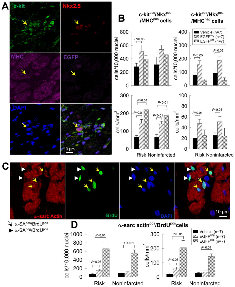
Figure 8. Cardiac commitment of endogenous CPCs after trasplantation of exogenous CPCs.
The commitment of endogenous CPCs (c-kitpos/EGFPneg cells) to cardiac lineage was assessed by quantitating endogenous CPCs that expressed Nkx2.5 and MHC in vehicle- and CPC-treated hearts. New myocytes formed in the last 2 weeks of life were identified by measuring α-sarcomeric actinpos and BrdUpos cells. (A) Representative confocal microscopic image of a c-kitpos/EGFPneg/Nkx2.5pos/MHCpos cell obtained in serial sections of a CPC-treated heart. (B) Quantitative analysis of c-kitpos/EGFPneg/Nkx2.5pos/MHCpos cells in the risk and noninfarcted regions of vehicle- and CPC-treated hearts (the CPC-treated group is subdivided into two subgroups, one with EGFPpos cells [EGFPpos] and one without EGFPpos cells [EGFPneg]). The number of c-kitpos/EGFPneg/Nkx2.5pos/MHCpos cells is normalized to both number of cells (104 nuclei, upper panels) and area (mm2, lower panels). Because cell density in CPC-treated, EGFPpos hearts was higher (due to presence of clusters of EGFPpos cells), the magnitude of the increase in endogenous CPCs in CPC-treated, EGFPpos hearts was greater when the number of cells was normalized to area (mm2) than to number of nuclei. (C) Representative confocal microscopic image of α-sarcomeric actinpos/BrdUpos cells in a CPC-treated heart. (D) Quantitative analysis of the number of α-sarcomeric actinpos/BrdUpos cells in the risk and noninfarcted regions of vehicle- and CPC-treated hearts (subdivided into EGFPneg and EGFPpos subgroups). Data are means ± SEM.

