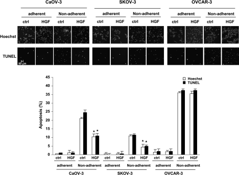
Figure 1.
HGF protected ovarian cancer cell from apoptosis induced by loss of cell adhesion. CaOV-3, SKOV-3, and OVCAR-3 cells were plated on plastic Petri dishes (adherent) or polyHEMA-coated dishes (nonadherent) in the presence or absence of 10 ng/ml HGF for 72 hours. Representative results from Hoechst staining (nuclear morphology) and TUNEL labeling were shown. Bar, 50 µm. The number of apoptotic cells were counted, and the bar diagram summarized the percentage of apoptotic cells from triplicate determination. Error bars indicate the SD of the mean. *P < .05 versus untreated controls.

