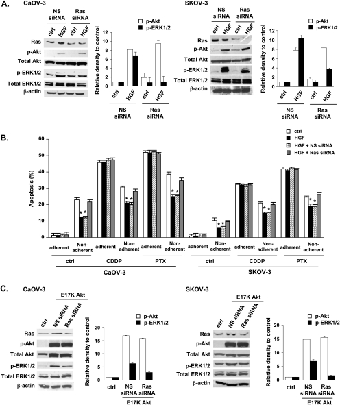
Figure 6.
PI3K functions upstream of Ras in response to HGF. CaOV-3 and SKOV-3 cells were transfected with non-specific (NS) siRNA or Ras siRNA for 24 hours and then treated with HGF (10 ng/ml) for 15 minutes. (A) Ras expression was determined by Western blot. Akt and ERK1/2 activities were analyzed using anti-phospho-specific (p-) Akt and p-ERK1/2 antibodies. (B) CaOV-3 and SKOV-3 cells plated on culture dishes (adherent) or polyHEMA-coated dishes (nonadherent) were transfected with NS siRNA or Ras siRNA for 24 hours and then subjected to 50 µM cisplatin (CDDP) or 100 nM paclitaxel (PTX) for 72 hours in the presence or absence of 10 ng/ml HGF. Cell apoptosis was determined with the TUNEL assay. The number of apoptotic cells was counted, and the bar diagram summarized the percentage of apoptotic cells from triplicate determination. (C) CaOV-3 and SKOV-3 cells were cotransfected with a constitutively active Akt (E17K) and NS siRNA or Ras siRNA for 24 hours. Ras expression was determined by Western blot. Akt and ERK1/2 activities were analyzed using anti-p-Akt and p-ERK1/2 antibodies. β-Actin was also included as a loading control. The signal intensity was determined by densitometry and expressed as p-Akt and p-ERK1/2 relative to total Akt and ERK1/2 for each sample. Error bar indicates the SD of the mean. *P < .05 versus untreated controls.

