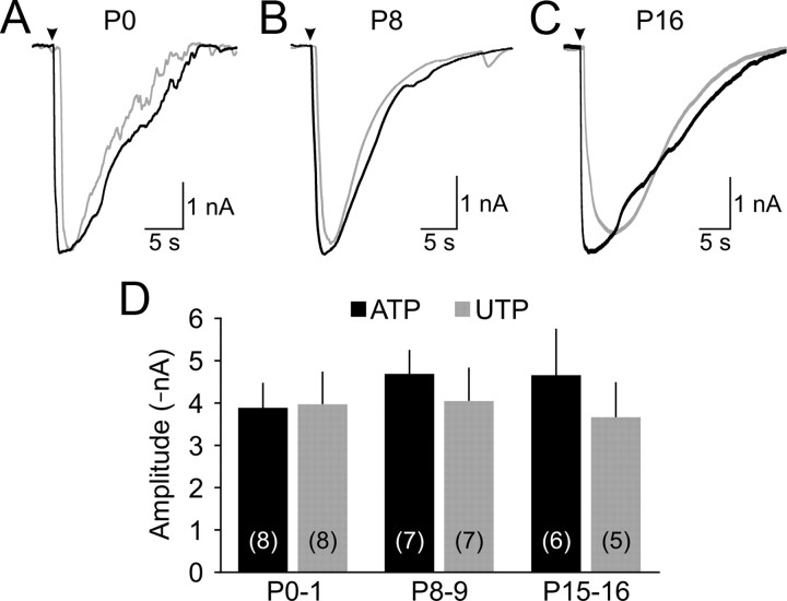
Figure 2.
Cochlear supporting cells remain responsive to ATP throughout early development. A–C, Representative inward currents elicited by local application (arrowhead) of ATP (100 μm; black traces) or UTP (100 μm; gray traces) to inner supporting cells in P0 (A), P8 (B), and P16 (C) cochleae. D, Graphs of average current amplitude (±SEM) evoked by ATP (black) or UTP (gray) at different postnatal ages. The number of experiments is indicated in parentheses.

