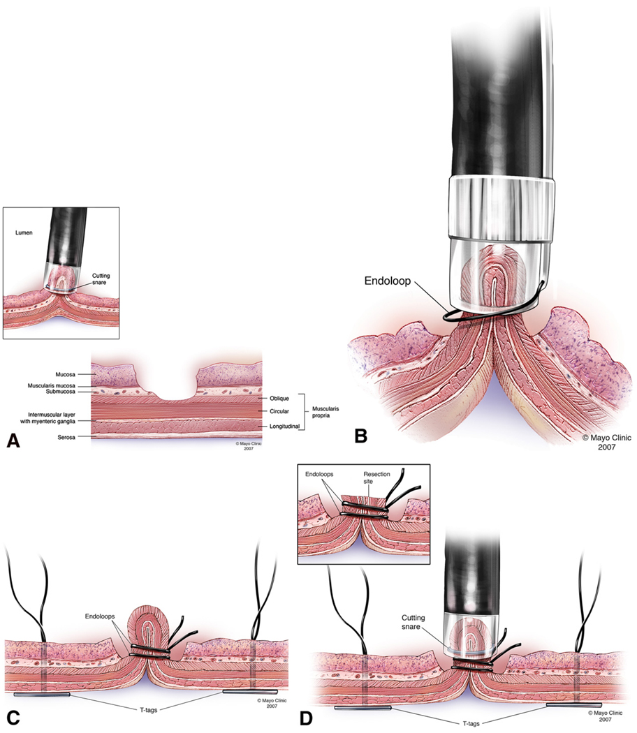
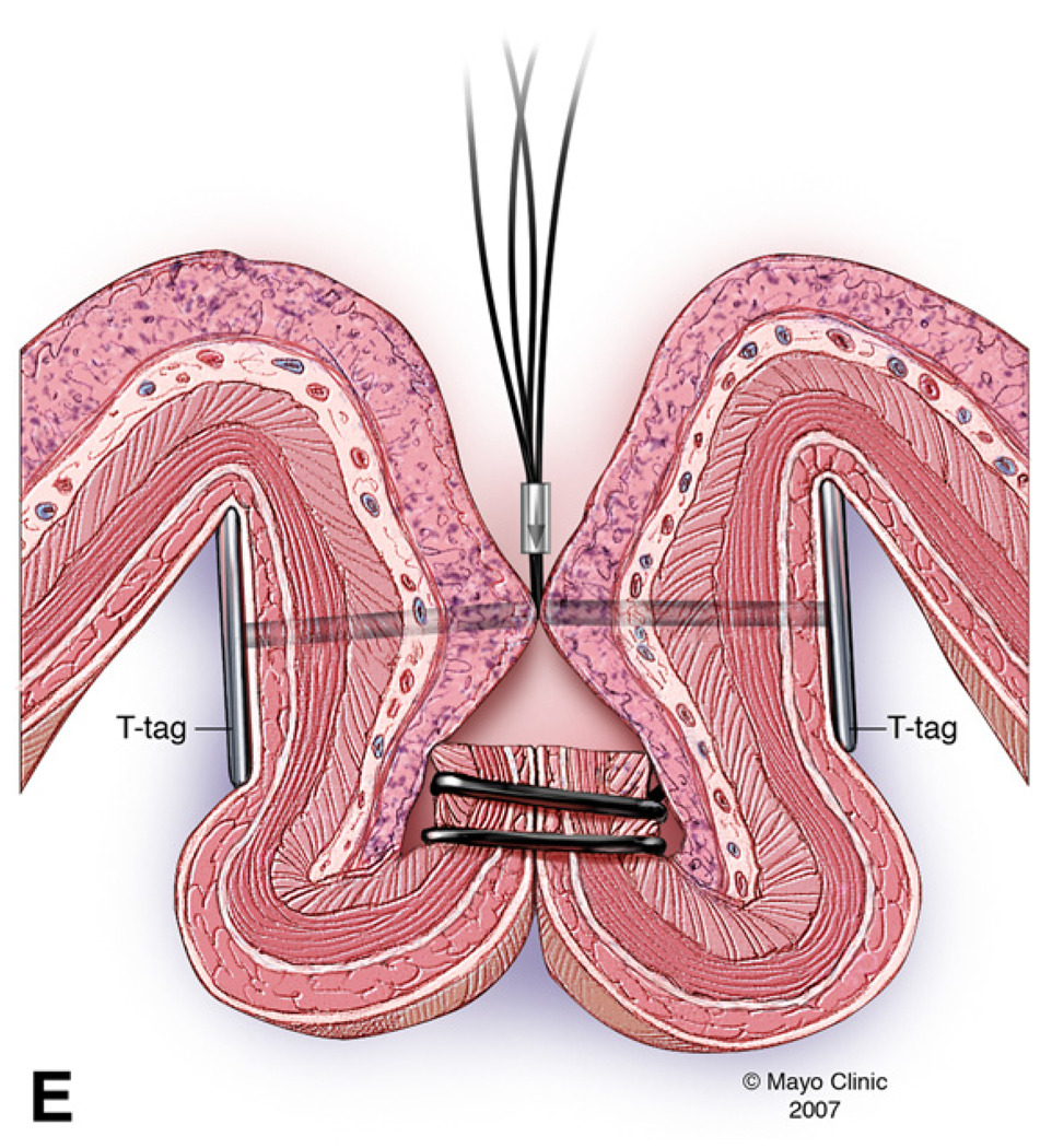
FIGURE 1.
FIGURE 1A: EMRC performed without a protective submucosal cushion
FIGURE 1B: Exposed muscle layer suctioned into EMRC cap while the endoloop was gently released and tightened around the pseudopolyp.
FIGURE 1C: Second endoloop and prototype T-tag tissue anchors are positioned around the base and adjacent to the pseudopolyp respectively.
FIGURE 1D: Pseudopolyp resected by snare electrocautery.
FIGURE 1E: Resection site further closed by tightening (opposing) prototype T-tag tissue anchors.


