Figure 1.
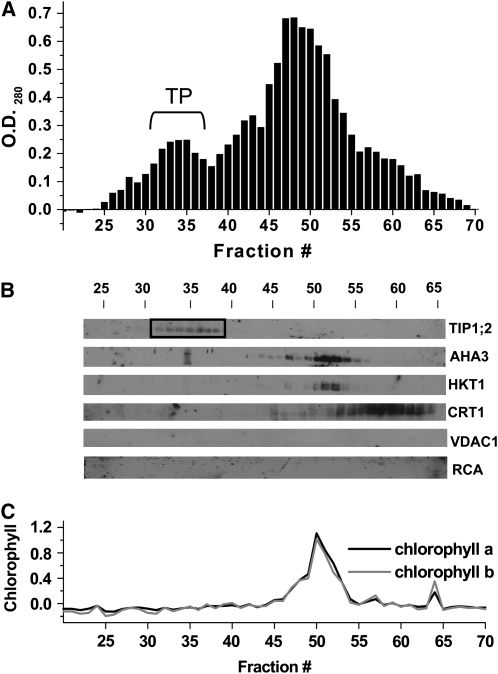
Purification of M. crystallinum Tonoplast by FFZE.
M. crystallinum microsomal membranes were separated by FFZE in the presence of 3 mM ATP.
(A) Protein profile of FFZE fractions showing a positive OD280. The bracket indicates the location of the ATP-dependent peak of tonoplast (TP).
(B) Immunological detection in the respective fractions of (from top to bottom) the tonoplast marker TIP1;2 (26 kD), the plasma membrane marker AHA3 (100 kD), the plasma membrane marker HKT1 (56 kD), the endoplasmic reticulum marker CRT1 (57 kD), the mitochondrial marker VDAC1 (29 kD), and the chloroplast marker RCA (43 and 41 kD). The fractions corresponding to pure tonoplast are enclosed in the box.
(C) Measurement of chlorophyll a and b in FFZE fractions.

