Figure 4.
PID-Independent GNOM Localization.
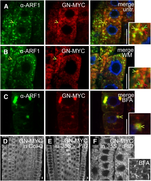
(A) to (F) Confocal images of anti-ARF1 and anti-MYC immunolocalizations. Bars = 10 μm.
(A) to (C) Partial ARF1 (green) and GNOM (GN-MYC) (red) colocalization in untreated seedlings (A) and seedlings treated with wortmannin (B) or BFA (C). Insets display enlarged regions highlighting colocalizing (yellow arrowheads) and noncolocalizing (red and green arrowheads) endosomes.
(D) and (E) Similar expression pattern of GNOM (GN-MYC under endogenous promoter) in the wild type (D) and 35SPro:PID (E).
(F) Normal subcellular distribution and response to BFA (BFA treatment is shown in the inset) of GNOM-MYC in 35SPro:PID-expressing seedlings. White arrowhead indicates GNOM accumulation in the BFA compartment.

