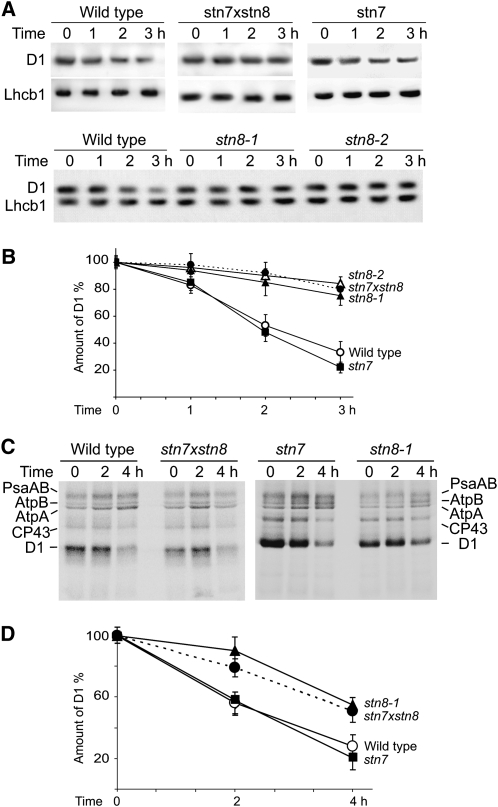
Figure 6.
Decreased D1 Turnover in stn7xstn8 and stn8.
(A) Immunoblot analysis of thylakoid proteins from wild-type, stn7, stn8-1, stn8-2, and stn7xstn8 plants with a D1-specific antibody and a control Lhcb1-specific antibody. Thylakoids were isolated from the leaves treated with lincomycin and exposed to high light of 900 μmol photons m−2 s−1 for the indicated periods of time.
(B) Time dependence of the D1 protein degradation in leaves of wild-type, stn7, stn8-1, stn8-2, and stn7xstn8 plants treated with lincomycin and exposed to high light like in (A). The values are means ± se of three independent experiments for each genotype.
(C) In vivo pulse-chase experiments with chloroplast proteins of wild-type, stn7, stn8-1, and stn7xstn8 plants labeled with [35S]Met and exposed to high light of 2000 μmol photons m−2 s−1. Four-week-old plants were labeled with [35S]Met for 2 h under dim light at room temperature in the presence of cycloheximide. After a 1-h chase period in dim light, including 30 min with lincomycin, plants were exposed to high light of 2000 μmol photons m−2 s−1 for 2 and 4 h, and proteins were analyzed by SDS-PAGE and phosphor imaging. Positions of the labeled subunits psaAB of photosystem I, AtpA and AtpB subunits of ATP synthase, and CP43 and D1 proteins of PSII are indicated.
(D) Time dependence of the labeled D1 protein degradation in leaves of wild-type, stn7, stn8-1, and stn7xstn8 plants subjected to in vivo pulse-chase experiments under high light as shown in (C). Amounts of labeled D1 protein were normalized relative to the sum of PsaAB, AtpA, AtpB, and CP43 bands. The values are means ± se of four independent experiments of each phenotype.

