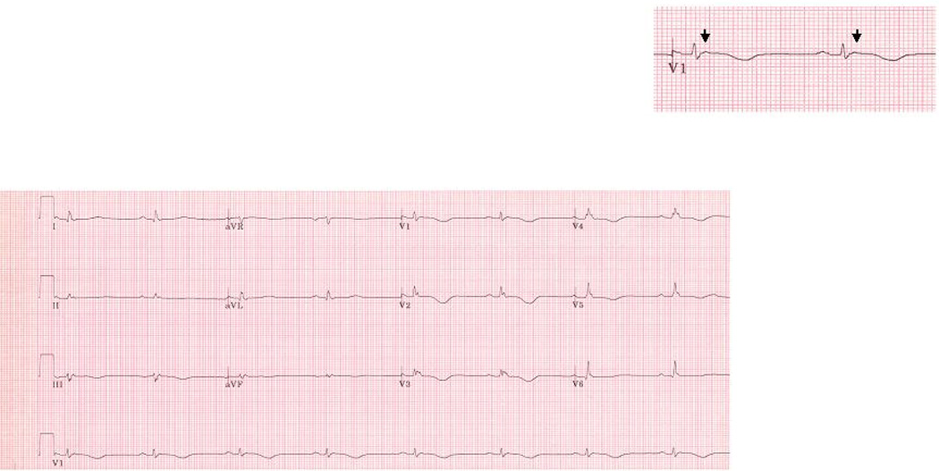
Figure 1.

Representative ECG obtained from an ARVD patient without IRBBB or CRBBB. This patient had an advanced form of ARVD. The arrow indicates an Epsilon wave. ECG also illustrates TWI in V1–V5, TAD ≥ 55ms in V1 and QRSd in V3 > 110 ms. This ECG also demonstrates low voltage, which in our experience is an uncommon finding in patients with ARVD and when seen is usually with advanced disease. There was no parietal block or localized right precordial QRS prolongation.
