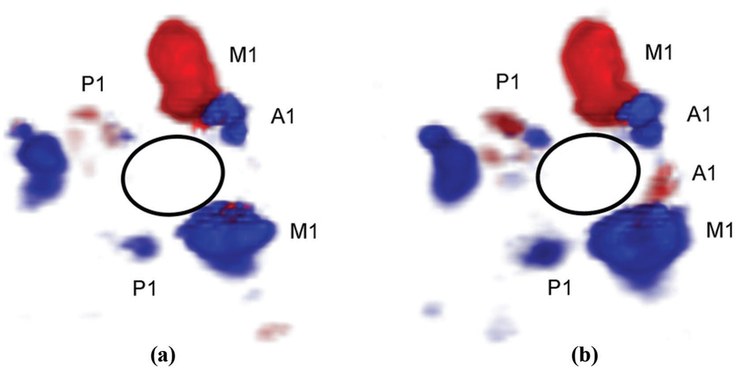
Fig. 1.
In vivo phase aberration correction: aberrated (a) and corrected (b) 3-D Doppler volume renderings of the circle of Willis, composed of ipsilateral and contralateral middle cerebral arteries (M1), posterior cerebral arteries (P1), and anterior cerebral arteries (A1). The corrected volume shows an increase in number of Doppler strength of 10% as well as the presence of the contralateral A1 not present in the aberrated volume.

