
FIGURE 1.
Case 26. Staining patterns (400× magnification): hematoxylin and eosin (top), CD3 immunostain (middle left), CD4 (middle right), CD25 (bottom left), and Foxp3 (bottom right). Note the lack of staining on the CD25 slide.
Official websites use .gov
A
.gov website belongs to an official
government organization in the United States.
Secure .gov websites use HTTPS
A lock (
) or https:// means you've safely
connected to the .gov website. Share sensitive
information only on official, secure websites.
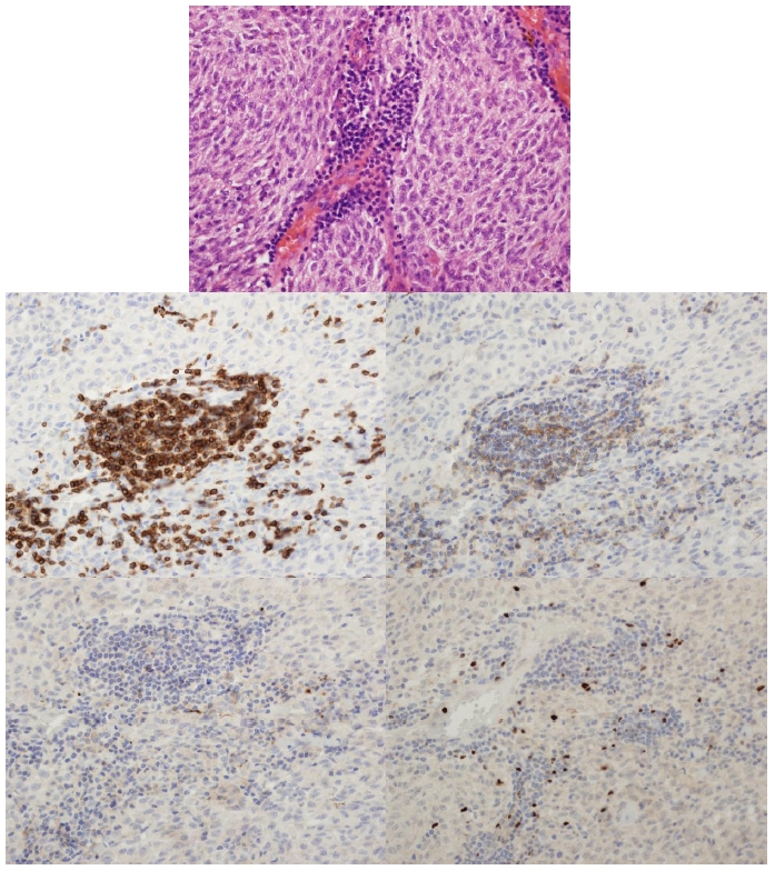
Case 26. Staining patterns (400× magnification): hematoxylin and eosin (top), CD3 immunostain (middle left), CD4 (middle right), CD25 (bottom left), and Foxp3 (bottom right). Note the lack of staining on the CD25 slide.