Figure 5.
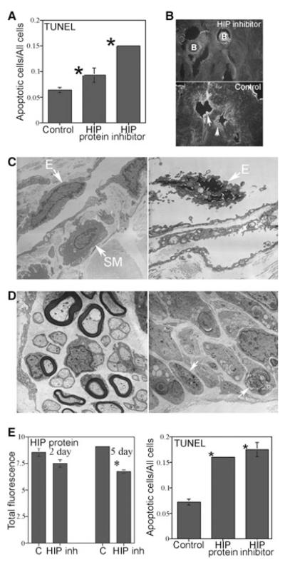
Affi-Gel beads soaked in hedgehog-interacting protein (HIP) inhibitor, HIP protein, and phosphate-buffered saline (control) were implanted under the pelvic ganglia of Sprague-Dawley rats and TUNEL, confocal microscopy, and HIP protein quantification were performed in the penis. (A) TUNEL assay showed that apoptosis was increased 2.4-fold in the corpora cavernosa after 2 days of HIP inhibition in the pelvic ganglia and 1.5-fold in the corpora cavernosa after HIP protein treatment of the pelvic ganglia. (B) Verification of HIP inhibitor function in vivo in the corpora cavernosa was performed by staining the HIP inhibited penis tissue with a second HIP antibody (Abcam ab39208). As the HIP inhibitor was already bound to HIP in the corpora cavernosal tissue, the Abcam antibody should not stain in the region of the Affi-Gel beads; however, staining should be present in untreated tissue away from the beads as was observed. Arrows indicate HIP protein. (X160). (C) Electron microscopy (EM) of HIP inhibited corpora cavernosa showed much less smooth muscle abundance by comparison with controls (×44,000) and endothelial apoptosis was evident (×70,000). (D) EM of HIP inhibited CN (×30,000) showed axonal degeneration and demyelination of nerve fibers by comparison with controls (×44,000). Arrows indicate degenerating axons. (E) HIP protein in the corpora cavernosa remained unchanged at 2 days of HIP inhibition in the pelvic ganglia but was significantly decreased 1.4-fold after 5 days of HIP inhibition in the pelvic ganglia. TUNEL assay showed that apoptosis was increased 2.4-fold in the corpora cavernosa after 2 days of HIP inhibition directly in the penis and 2.2-fold in the corpora cavernosa after direct HIP protein treatment of the penis via Affi-Gel beads. B = Affi-Gel bead; E = endothelium; SM = smooth muscle.

