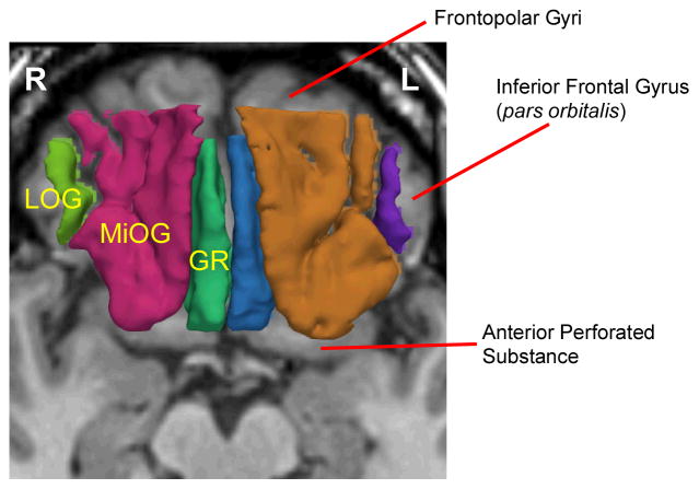
Figure 2.
MR Images of Three Orbitofrontal Subregions. 3D reconstruction of the three orbitofrontal subregions of Gyrus Rectus (GR; left: blue, right: green), Middle Orbital Gyri (MiOG; left: brown, right: red), and Lateral Orbital Gyrus (LOG; left: purple, right: light green), superimposed on axial plane of SPGR image.

