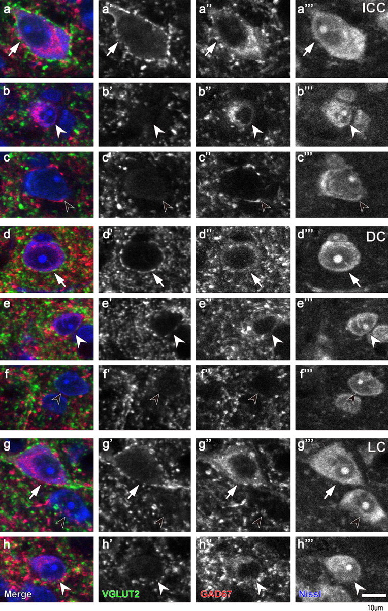
Figure 3.

Confocal images that show two types of neurons immunopositive for GAD67 (red). a, d, g, Immunopositive cells with dense VGLUT2+ axosomatic endings (green) are indicated with arrows. b, e, h, GAD67+ cells without VGLUT2+ terminals (white arrowheads). c, f, g, GAD67-negative cells (black arrowheads) also lack VGLUT2+ axosomatic endings. These three types were seen in all IC subdivisions (a–c, ICC; d–f, DC; g and h, LC). Scale bar: (in h′′′) a–h′′′, 10 μm.
