
Figure 4.
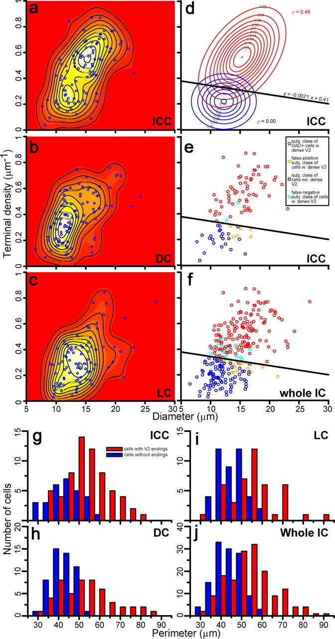
Analysis of GAD67+ IC cells based on cell diameter and density of VGLUT2+ axosomatic endings and comparison to subjective classification. a–c, Scatter plots of GAD67+ cells (blue circles) in subdivisions of the IC and kernel density estimates of populations. Kernel density estimates are visualized by both color depth (white and red indicate high and low probability density, respectively) and contour plots. d, Best fit of bivariate Gaussian distribution to the kernel density estimate from the ICC with two bivariate normal distributions calculated by the least-squares method. The distribution with lower terminal density (blue contours) shows no covariance (ρ = 0) between diameter and density, while the distribution with higher terminal density (red contours) shows covariance between these parameters (ρ = 0.49). Points at which both distributions have the same probability density aligned with a slope y = −0.0071x + 0.41. e, Scatter plot of the ICC GAD67+ cells (circles, 92 cells) is divided by slope (y) into two populations. Neurons were classified subjectively, as VGLUT2-axosomatic positive (red, 61 cells) or VGLUT2-axosomatic negative (dark blue, 23 cells). A few cells were miscategorized and represent false-positive (cyan, 2 cells) and false-negative (orange, 6 cells) choices. See also Table 1. f, Scatter plot of all GAD67+ cells in all IC subdivisions classified by our subjective analysis and separated by slope (y) used in d and e. VGLUT2-axosomatic positive (140 cells; red circles) and VGLUT2-axosomatic negative (98 cells; blue circles). Errors were 21 false-positive and 20 false-negative cells. See also Table 2. g–j, Histograms of perimeter show the distribution of GAD67+ cells with or without VGLUT2+ axosomatic endings (red and blue bars, respectively) classified objectively in the ICC (g), DC (h), LC (i), and entire IC (j). In all cases, these two cell types make two distinct but overlapping populations of GAD67+ neurons.