
Fig. 1.
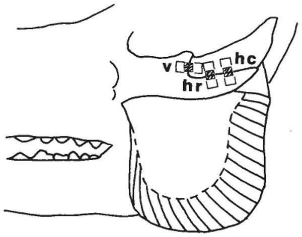
Diagram of the zygomatic arch and masseter muscle of the pig, showing the location of uniaxial strain gages placed over the zygomatico-squamosal suture. Note differing muscle fiber orientations along the rostrocaudal axis of the masseter. The gage elements were aligned along the long axis of each rectangle. The hatched area in the center of each rectangle shows the area (underlying the gage element) which was not bonded to the underlying tissue. v = vertical segment; hr = horizontal segment, rostral portion; hc = horizontal segment, caudal portion.