
Fig. 3.
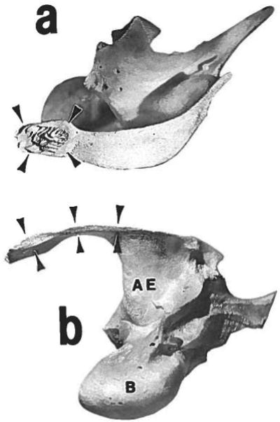
Disarticulated left squamosal bone, seen in lateral aspect (a) and inferior aspect (b). Rostral is to the left. The sutural articular surfaces are marked with arrowheads. The surface for the vertical segment is seen in the lateral view (a). Note the chevron pattern of bony laminae; these interdigitate with similar laminae on the medial side of the zygomatic bone. The surface for the horizontal segment is seen in the inferior view (b); it is nearly smooth, as is the superior surface of the zygomatic bone at this location. AE = articular eminence; B = auditory bulla.