
Fig. 5.
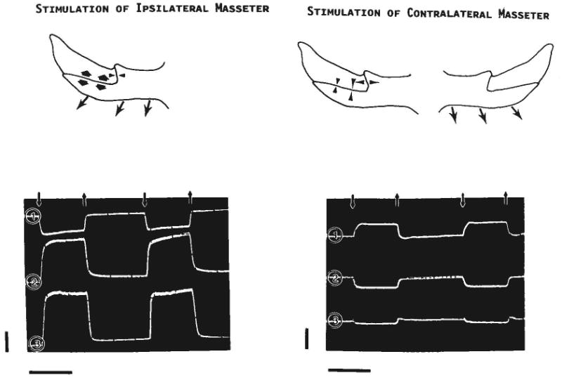
Results of stimulating the ipsilateral (left) or contralateral (right) masseter. The drawings depict strains (arrowheads) seen in the right zygomatico-squamosal suture when the masseter (arrows) contracts. The lower tracings were photographed from the oscilloscope screen (pig 105). Downward small arrows indicate the onset of the stimulus, upward small arrows its offset. Channel 1 is from the gage on the vertical segment of the suture and shows compression (negative values) during ipsilateral stimulation and tension (positive values) during contralateral stimulation. Channels 2 and 3 are, respectively, from the gages at the rostral and caudal locations along the horizontal segment; both show tension during ipsilateral stimulation and compression during contralateral stimulation. Calibration bars: vertical = 1,000 microstrain; horizontal = 1 second.