
Fig. 8.
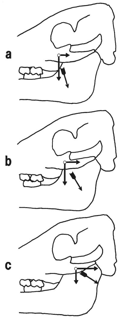
Interpretation of the effect of partial masseter contraction on strain in the zygomatico-squamosal suture. a: Contraction of rostral fibers, such as occurs during simple closing movements of the mandible (e.g., pig 105) or on the balancing side in unilateral mastication (e.g., left side in pig 104). Caudal and ventral components of the muscle force are shown acting on the zygomatic bone. The horizontal component, assumed to be responsible for compression in the vertical segment of the suture, is relatively small. The ventral component, assumed to be responsible for tension in the horizontal segment of the suture, is large, but it is distant from the horizontal segment. Thus strains are low in both parts of the suture. b: Contraction of middle fibers. These fibers represent the average pull of the whole muscle; thus strains produced are similar to those produced by the masseter on the working side during mastication (e.g., right side of pig 104; pig 107). Both caudal (compressing the vertical segment) and ventral (tensing the horizontal segment) components of the muscle force are large, c: Contraction of caudal fibers, seen when the working side masseter moves the mandible toward the balancing side at the end of the power stroke of mastication. The caudal component of force is large, while the ventral component is small, accounting for high compressive strain in the vertical segment of the suture but low tensile strain in the horizontal segment.