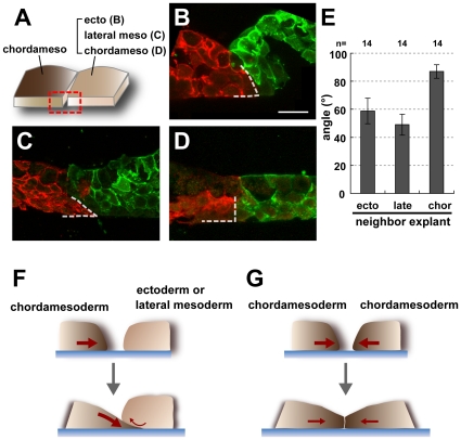
Figure 3. Geometry of the vertical section of the explants in the conjugation assay.
(A) Diagram showing the area observed by section. The red box indicates the observed area, which is the vertical plane of the boundary between the chordamesoderm and the contiguous tissue. (B, C, D) Vertical sections of the chordamesoderm (red) and its neighboring tissue (green). The chordamesodermal tissue had a wedge-like shape (dotted lines)when conjugated with ectoderm (B) and lateral mesoderm (C), but the edge of the tissue remained essentially vertical in homogeneous conjugation assays (D). (E) Graph showing the average angle of chordamesodermal tissues conjugated with neighboring tissues. (n = 14 in each group, ecto: ectoderm, late: lateral mesoderm, chor: chordamesoderm) (F, G) Model for the creation of the boundary geometry. The chordamesodermal tissue crawled under the ectoderm or lateral mesodermal tissues because of its strong adhesion to the fibronectin, creating a wedge-like shape, and attachment of the apical side of the chordamesodermal cells to the neighboring explant (F). On the other hand, the orthogonal-like shape was formed because of the cells' equal adhesion to the fibronectin when chordamesoderm was conjugated to itself (G).

