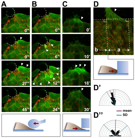
Figure 4. Mechanical stimuli change the intracellular calcium dynamics and cell polarity.
(A, B) Increase in the intracellular calcium in the chordamesodermal tissue by glass needle pushing. The dotted lines show the edge of the glass needle. Before the pushing of the glass needle (0 sec), the calcium did not show any activated signals. Just after the pushing of the needle (9 sec in A, 6 sec in B), the cells responded to the stimulus by an increase in intracellular calcium (arrowheads). The calcium propagation was either restricted to a single cell (A), or to several cells (B), and disappeared within a minute. (C) Increase in intracellular calcium (arrowheads) in the chordamesodermal tissue by cells crawling underneath a block of silicone. (D) Cell alignment after silicone block attachment. One hour after the attachment in (C), the cells behind the crawling cells (arrowhead) aligned perpendicular to the silicone block (area indicated by a). The horizontal dotted line indicates the edge of the silicone block. (D′, D″) Rose diagrams showing the distribution of the cells' angles in D. The cells behind the cells that received the stimulus (a in D) aligned almost perpendicular to the edge of the silicone block (D′), but cells that did not receive the stimulus (b in D), did not (D″).

