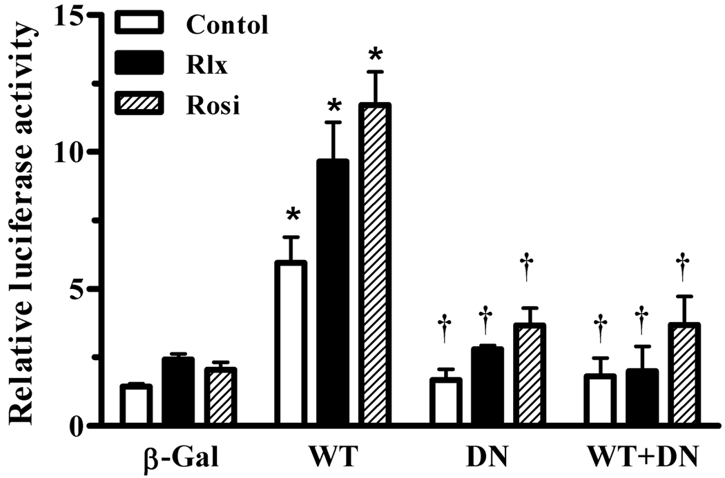
Fig. 3.
Effect of relaxin on wild-type and dominant-negative PPARγ. HEK-RXFP1 cells were infected with adenoviruses expressing β-galactosidase (β-Gal), wild-type PPARγ (WT), dominant-negative PPARγ (DN) or WT:DN at a 1:2.5 ratio. Cells were then treated with relaxin (1 nM), rosiglitazone (1 µM), or vehicle for 24 hours, then subject to the PPRE luciferase assay. The data are expressed as the ACO-PPRE luciferase activity relative to that in uninfected untreated cells. Data are mean ± S.E.M., N=3. *p<.001 compared to β-Gal. †p<.01 compared to WT.

