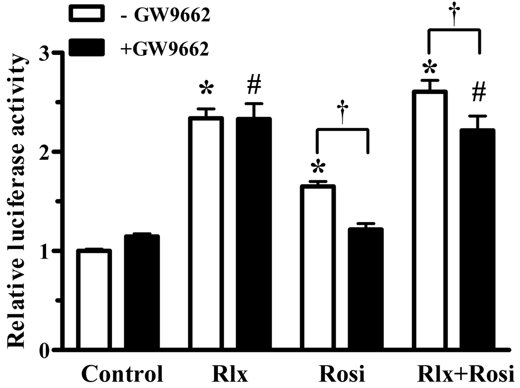
Fig. 7.
Relaxin activation of PPARγ is not ligand-dependent. HEK-RXFP1 cells were treated for 24 hours with 1nM relaxin (Rlx), 1µM rosiglitazone (Rosi), or both, in the presence and absence of the inhibitor of PPARγ ligand binding (GW9662, 100nM) or vehicle control (DMSO) for 24 hours, then subject to the PPRE-luciferase assay. The data are expressed as the ACO-PPRE luciferase activity relative to that of untreated cells, mean ± S.E.M, N=4. *p<.001 compared to untreated controls; #p<.01 compared to GW9662 alone; †p<.05.

