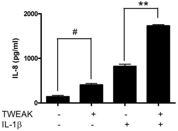
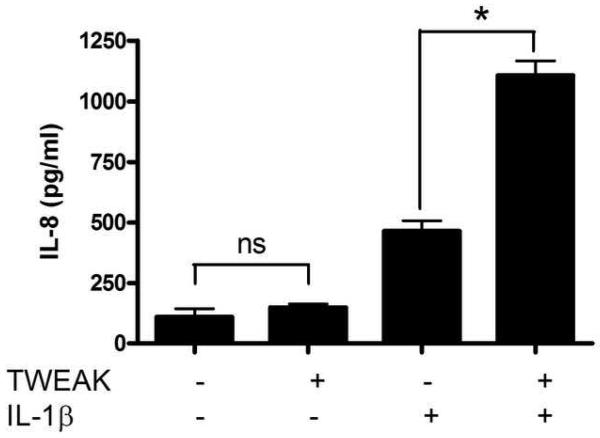
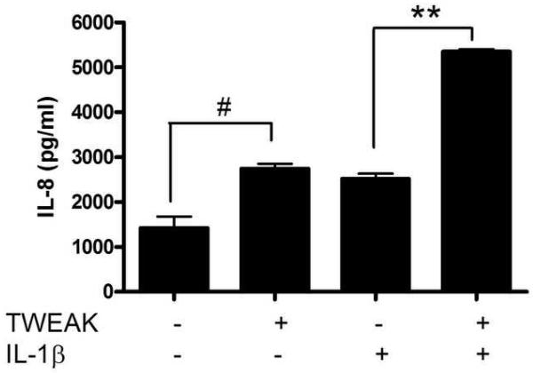
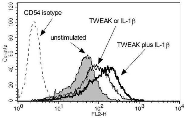
Figure 4. TWEAK induces the secretion of IL-8 by vaginal and cervical epithelial cells and potentiates the effect of IL-1ß.
Immortalized vaginal (A), ectocervical (B), and endocervical (C) epithelial cells were stimulated with 1 μg TWEAK per ml, 5 ng IL-1ß per ml, or both, for 24 hours. Culture supernatants were harvested and assayed for IL-8 by ELISA. The values are derived from IL-8 standard curves and represent the mean ± the SEM from triplicate wells, and are representative of at least three independent experiments. (D) Immortalized vaginal epithelial cells were stimulated with 1 μg TWEAK per ml, 5 ng IL-1ß per ml, or both, for 24 hours. Cells were harvested, stained for surface expression of CD54, and analyzed by FACS. The vertical axis represents the relative cell number, while the horizontal axis represents the intensity of anti-CD54 staining. Basal expression of CD54 is shown in the gray shaded histogram. Treatment with TWEAK alone is shown in the thin line while treatment with IL-1ß alone is in the dashed lines. Treatment with TWEAK plus IL-1ß is shown in the thick line. Surface staining with the isotype control antibody for unstimulated cells is shown in the gray dashed line, and stimulation of the cells had no effect on isotype staining (data not shown). Similar results were found with the ectocervical epithelial cells (data not shown). These data are representative of two independent experiments.
* p<0.001; ** p<0.0001 for IL-1ß alone vs. TWEAK + IL-1ß.
# p<0.01 for unstimulated vs. TWEAK alone. ns, not significant .


