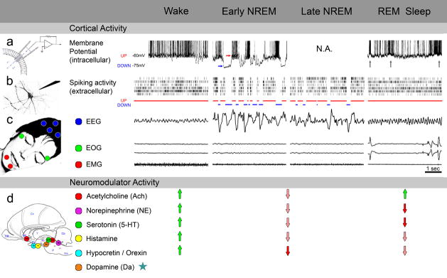
Figure 2. Neurophysiology of wake and sleep states.
A comparison of cortical activity (upper panel) and neuromodulator activity (bottom panel) in wake, early NREM (when sleep pressure is high and dream reports are rare), late NREM (when sleep pressure dissipates, and dream reports are more frequent), and REM sleep (when dreams are most common).
(a) Intracellular studies. The membrane potential of cortical neurons in both wake and REM sleep is depolarized and fluctuates around −63mV and −61mV, respectively [77]. In REM sleep, whenever phasic events such as rapid eye movements and PGO waves occur (gray arrows, events not shown), neurons increase their firing rates to levels that surpass those found in wake [77, 146]. In early NREM sleep, neurons alternate between two distinct states, each lasting tens/hundreds of milliseconds: UP states (red arrow) are associated with depolarization and increased firing, while in DOWN states (blue arrow) the membrane potential is hyperpolarized around −75mV, and neuronal firing fades[78, 147]. Intracellular studies focusing specifically on late NREM sleep are not available (N.A.).
(b) Extracellular studies. Spiking of individual neurons in REM sleep reaches similar levels as in active wake. In both wake and REM sleep, neurons exhibit tonic irregular asynchronous activity [77, 148–151]. Sustained activity in wake and REM sleep can be viewed as a continuous UP state [78] (red bars). In early NREM sleep, UP states are short and synchronous across neuronal populations, and are frequently interrupted by long DOWN states (blue bars). In late NREM sleep, UP states are longer and less synchronized [79].
(3) Polysomnography. Waking is characterized by low-amplitude, high-frequency EEG activity (above 7Hz), occasional saccadic eye movements, and elevated muscle tone. In early NREM sleep, high-amplitude slow waves (below 4Hz) dominate the EEG. Neuronal UP (red) and DOWN (blue) states correspond to positive and negative peaks in the surface EEG, respectively [79]. Eye movements are largely absent and muscle tone is decreased. In late NREM sleep, slow waves are less frequent, while spindles (related to UP states and surface EEG positivity) become more common. Eye movements and muscle tone are largely similar to early NREM sleep [152]. In REM sleep, theta activity (4–7 Hz) prevails, rapid eye movements occur, and muscle tone is dramatically reduced.
(d) Neuromodulator activity. Subcortical cholinergic modulation is highly active in wake and REM sleep (green arrows) and leads to sustained depolarization in cortical neurons and EEG activation [77]. Wake is further maintained by activity of monoamines, histamine, and hypocretin/orexin (green arrows). In sleep, monoaminergic systems including norepinephrine and serotonin reduce their activity (pink arrows), and are silent in REM sleep (red arrows). While dopamine levels do not change dramatically across the sleep-wake cycle (asterisks), phasic events and regional profiles may differ[153].
Data are pooled across different species for illustration purposes. Intracellular cat data adapted with permission from Ref [77]; extracellular and EEG rat data obtained from V. Vyazovskiy (personal communication).

