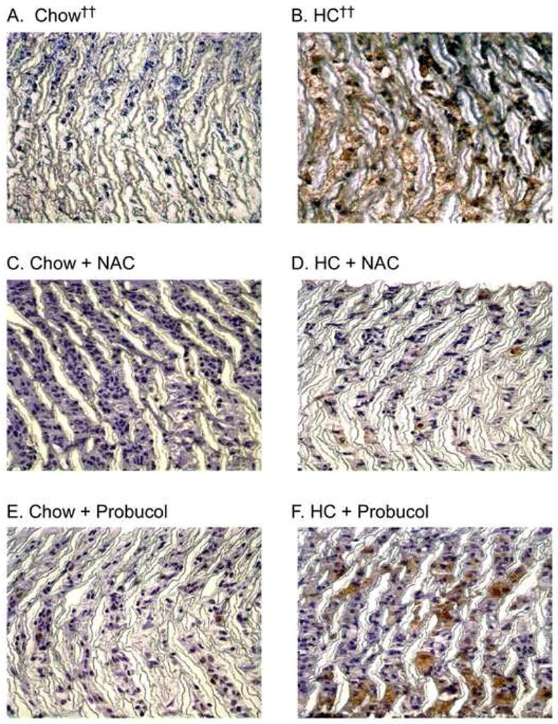
Fig. 4.

Macrophage infiltration. Following perfusion-fixation with 4% paraformaldehyde, graft sections were processed for immunohistochemistry and stained using mouse anti-rabbit macrophage antibody (RAM-11). Representative sections are shown from each rabbit group (A) chow diet (n = 6), (B) high cholesterol diet (HC, n = 5), (C) chow plus NAC (n = 3), (D) HC plus NAC (n = 4), (E) chow plus probucol (n = 5), (F) HC plus probucol (n = 5). Original magnification ×100. †† Data from reference 10
