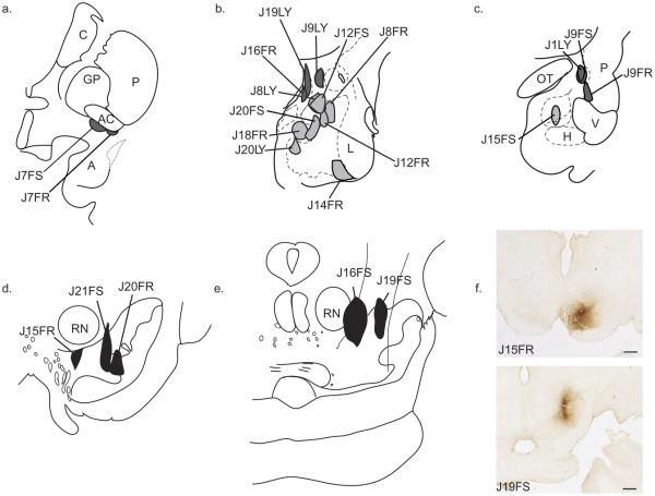
Fig. 2.
Schematic of all injection sites. Retrograde injections (a–c): (a) Injection sites in the IPAC (J7FR, J7FS). (b) Injection sites in the “amygdala proper” are colored light gray: Bmc (J8FR, J12FR); ABmc (J8LY, J12FS); ABpc (J18FR, J20FS); Bpc (J14FR, J20LY). Injections sites in the “extended amygdala” are colored dark gray: CeM (J9LY); MeN (J16FR, J19LY). (c) Injection sites in the caudal “amygdala proper”: Bpc (J15FS). Injection sites in caudal extended amygdala: CeLcn (J1LY, J9FS); Amygdalostriatal area (J9FR). Anterograde injections (d–f): (d) Injection sites in the A9-SNpc: medial A9-SNpc (J15FR); mediolateral A9-dt and A9-vt (J21FS); medial A9-vt (J20FR). (e) Injections sites in the A8-RRF: rostromedial A8-RRF (J19FS); caudomedial A8-RRF (J16FS). (f) Photomicrographs of injection sites: J15FR—medial A9-SNpc (top); J19FS—rostromedial A8-RRF (bottom). FR, Fluoro Ruby; FS, Fluorescein; LY, Lucifer Yellow. Scale bar=1 mm. For interpretation of the references to color in this figure legend, the reader is referred to the Web version of this article.

