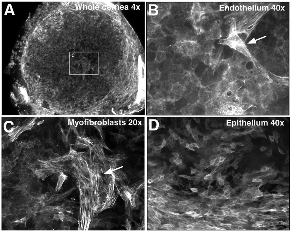
Figure 5.
Abnormal expression of α-SMA in TGFβ1 transgenic mouse cornea at postnatal day 16. Whole-mount immunofluorescent staining for α-SMA and images were analyzed by using confocal microscopy. α-SMA-expressing cells were present in both the corneal endothelial (A–C) and epithelial (D) layers of the TGFβ1 transgenic mouse. Cells with a myofibroblast shape and a greater level of α-SMA were found in the corneal endothelial layer (as indicated by the arrows in B and C).

