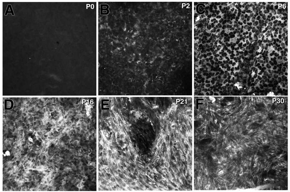
Figure 6.
Increase of αSMA expression along with the progression of myofibroblast formation in the postnatal TGFβ1 transgenic cornea. α-SMA expression was not detected in the transgenic cornea at postnatal day (P) 0 (A). A few α-SMA-positive cells were found at P2 (B) and the number of α–SMA-expressing cells increased significantly between P6 to P30 (C–F). The myofibroblast-like morphology became visible in the transgenic cornea at P16 (D). By P30 almost all of the αSMA-positive cells in the transgenic cornea were transformed into myofibroblast phenotypes including irregular shape, and the presence of stress fibers.

