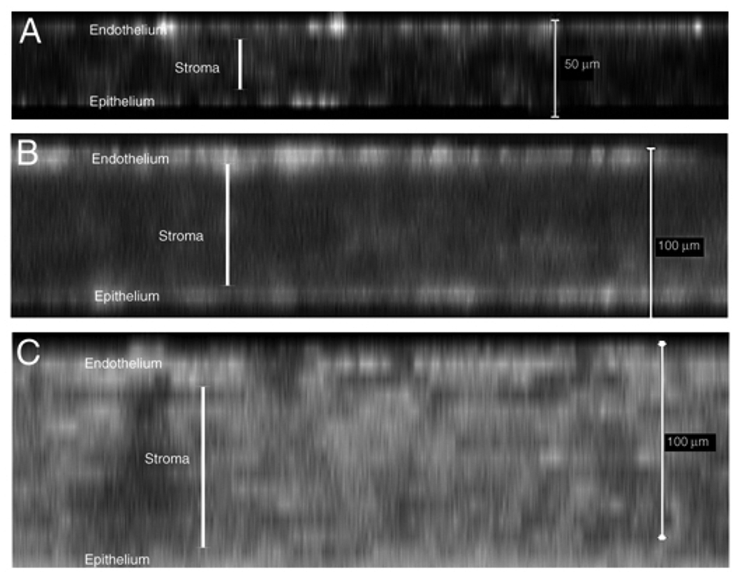
Figure 7.
Distribution patterns of αSMA-expressing cells in the cornea of the TGFβ1 transgenic mice. Confocal x–z images of whole-mount-stained corneas shown at different postnatal ages. At P7 (A) and P16 (B), most of the αSMA-expressing cells were localized in the corneal epithelial and endothelial layers. By P30 (C), strong α-SMA staining was distributed in all corneal cell layers. Cornea edema along with the enhanced expression of α-SMA progressed in the transgenic corneas from P7–P30.

