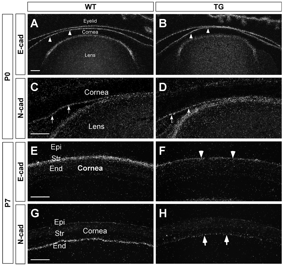
Figure 8.
Decrease in cadherin expression in the cornea of the TGFβ1 transgenic mouse. At P0 (A–D), E-cadherin was expressed in the corneal epithelial cells (arrowheads) and N-cadherin was in the corneal endothelial cells (arrows). E-cadherin expression was also detected in the epithelial cells of the eyelid and lens, and N-cadherin in the lens epithelial cells. Similar E- and N-cadherin expression patterns were found for the wild-type and the TGFβ1 transgenic mice, suggesting that prenatal development of the cornea and lens were not affected in the OVE853 transgenic mice. At P7 (E–H), E- and N-cadherin expression decreased in the corneal epithelial (arrowhead in F) and endothelial layers (arrows in H), respectively, in the TGFβ1 transgenic mouse, a feature consistent with EnMT and EMT in the cornea of the transgenic mouse. Scale bar represents 100 µm.

