
Figure 9.
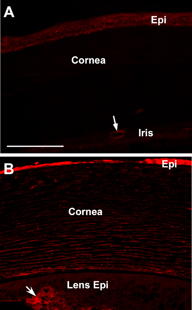
Cadherin-11 immunofluorescence in P21 mouse cornea. Cadherin-11 staining was found in the vescular tissue of the iris (arrow in A). A low level of cadherin-11 expression was also detected in the WT corneal epithelial layer (A). The intensity of cadherin-11 staining was increased in the transgenic mouse cornea, particularly in the corneal epithelial layer (B). On the same section, cadherin-11 was also shown in the group of lens epithelial cells (arrow in B) which were positive for αSMA and had transformed into myofibroblasts.