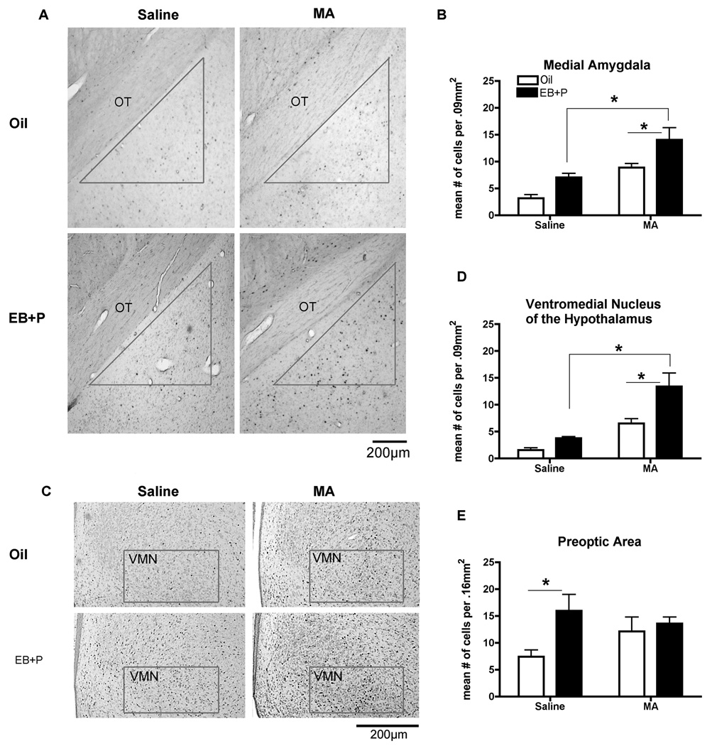
Figure 6.
Effect of ovarian hormones and MA on Fos-immunoreactivity (ir) in the (A) the Medial Amgydala and (B) Ventromedial Nucleus of the Hypothalamus. Adult ovariectomized Sprague-Dawley rats were primed with EB 48 h (5µg) and 24 h (10µg) prior to the day of collection. Four hours before the collection, animals received an injection of oil vehicle or 500µg P. MA (5mg/kg) was co-administered on each day of the hormonal priming. The photomicrographs represent the Fos-ir in the medial amygdala (A) and the ventromedial nucleus of the hypothalamus (B). The contour represents the counting area used to quantify the number of Fos-positive cells. OT, optic tract. Scale bar: 200 µm. A two-way ANOVA followed by Bonferroni t-tests indicated the combination of EB+P and MA significantly increases Fos-ir compared to either oil control and MA treatments or EB+P and saline controls (*p<0.05) in both brain regions. (n = 7 to 8 animals in each group). (C) Effect of ovarian hormones on Fos-ir in the medial preoptic area. A two-way ANOVA followed Bonferroni t-tests indicated that EB+P and saline-treatment significantly increased Fos-ir compared to the oil and saline-control (*p<0.05). Data are represented as means ± SEM. (n = 7 to 8 animals in each group).

