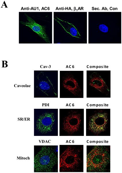
Figure 1. Location of AC6 and ß1AR transgene proteins.
A. Immunofluorescence staining and deconvolution analysis of cardiac myocytes after gene transfer of Ad.AC6 and Ad.ß1AR. Uninfected cardiac myocytes served as a control (Con). Anti-AU1 antibody was used to detect AC6 transgene (green), anti-HA for ß1AR transgene (green), and Hoechst dye was used to identify the nucleus (blue). AC6 transgene was evenly distributed in the plasma membrane and cytoplasm, but not within the nucleus. In contrast, ß1AR transgene was limited primarily to the plasma membrane (40X).
B. Double immunofluorescence staining of AC6 transgene by anti-AU1 antibody (red) with anti-caveolin 3 (Cav-3) antibody (green, for caveolae); with anti-voltage dependent anion selective channel protein (VDAC) antibody (green, for mitochondria); and with anti-protein disulphide-isomerase (PDI) antibody (green, for sarcoplasmic reticulum). AC6 transgene was localized in caveolae, mitochondria and SR.

