
Figure 2. Sagittal and coronal MRIs confirming placement of guide cannula above the dorsal putamen.
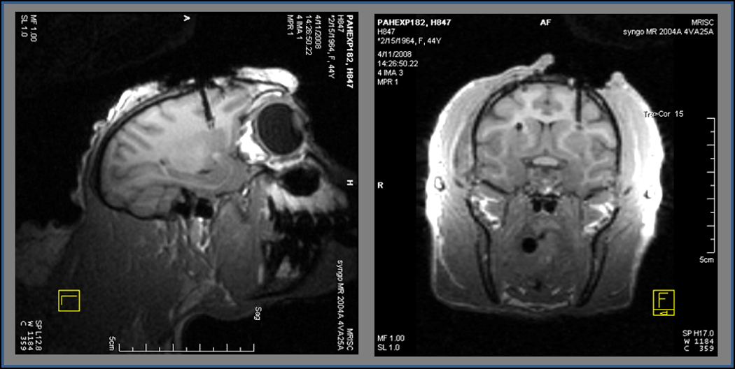
Three-dimension anatomical T1-weighted images were collected on a 3T Siemens Trio clinical imager to confirm correct placement of the guide cannula. Through the cannula, the SG-2 MEA was targeted to the dorsal putamen for amperometric glutamate measurements in NHPs.