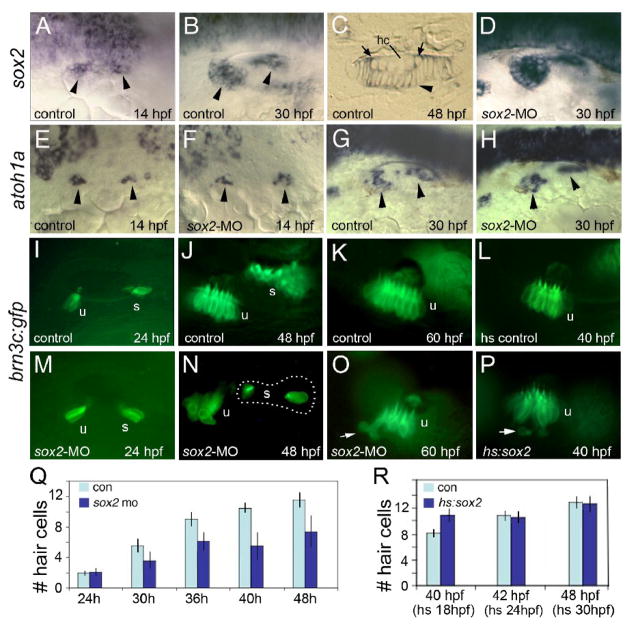
Figure 1. Sox2 is not required for hair cell development.
(A–C) sox2 expression in control embryos at 14 hpf (A), 30 hpf (B) and in a cross section of the utricular macula at 48 hpf (C). sox2 expression is lost from mature hair cells (hc) but is still detected in recently formed hair cells (arrows) and all surrounding support cells (arrowhead). (D) sox2 expression at 30 hpf in a sox2 morphant. (E–H) Expression of atoh1a in control embryos (E, G) and sox2 morphants (F, H) at the indicated times. Arrowheads mark macular expression domains. (I–P) brn3c:gfp expression in control embryos at 24 hpf (I), 48 hpf (J) and 60 hpf (K); expression in a control embryo heat shocked at 24 hpf and photographed at 40 hpf (L); expression in sox2 morphants at 24 hpf (M), 48 hpf (N) and 60 hpf (O); and expression in a hs:sox2 transgenic embryo heat shocked at 24 hpf and photographed at 40 hpf (P). Positions of the utricular (u) and saccular (s) maculae are indicated. Note the absence of hair cells in the middle of the saccular macula in the sox2 morphant (N). Arrows in (O, P) show hair cells being extruded from the utricular macula. All images show lateral views with anterior to the left and dorsal to the top. (Q) A time course showing the mean number of utricular hair cells in control embryos (con) and sox2 morphants (sox2 mo). Sox2 morphants exhibited a normal number of hair cells at 24 hpf (p = 0.88) but showed significantly fewer hair cells at later time points (p < 0.0001 for each time point). (R) Number of utricular hair cells in control embryos and hs:sox2/+ embryos subjected to heat shock at 18, 24 or 30 hpf, and counted at 40, 42 or 48 hpf, respectively. Transgenic embryos heat shocked at 18 hpf produced significantly more hair cells than normal (p < 0.0004), whereas the number of hair cells was not altered by heat shocking at 24 or 30 hpf (p = 0.78 or 0.73, respectively). Error bars in (Q, R) represent standard deviations, with n ≥ 15 for each time point.

