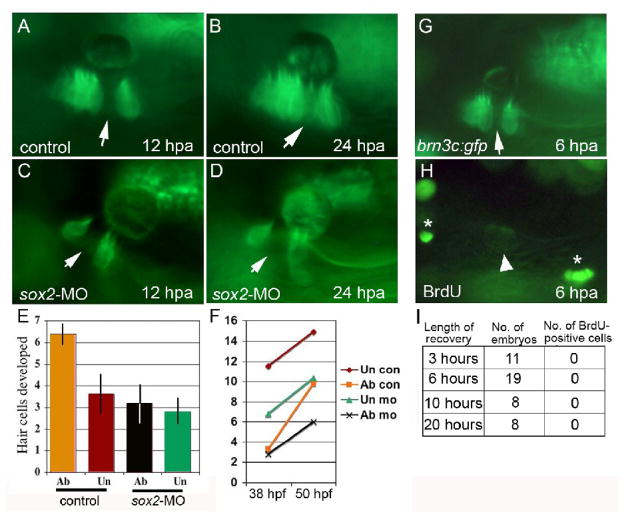
Figure 4. Hair cell regeneration requires sox2 but does not involve cell division.
(A–D) brn3c:gfp following ablation in a control embryo (A, B) and a sox2 morphant (C, D). Hair cells were ablated at 48 hpf, and ablated regions (arrows) were still evident at 12 hours post-ablation (hpa) (A, C) and 24 hpa (B, D). By 24 hpa, the gap filled in with newly formed hair cells in the control (B) but not in the sox2 morphant (D). (E, F) The number of hair cells produced following wholesale ablation of utricular hair cells. Ablation was conducted at 30 hpf, embryos were allowed to recover, and hair cells were counted at 38 hpf and again at 50 hpf. Typically 2 hair cells were produced during the recovery period. The number of hair cells produced between 38 and 50 hpf (E), and the total number of hair cells (F) are indicated for ablated (ab) and unablated (un) control embryos and sox2-morphants. Each time point shows the mean ± standard error of 3 or 4 experiments, with sample sizes of 19 to 23 embryos. (G–I) BrdU incorporation at various times following ablation initiated at 48 hpf. After 3, 6, 10 or 20 hours of recovery, embryos were incubated with BrdU for 3 hours and then fixed for processing. A specimen just before fixation at 6 hours post ablation (G) shows that the hair cell gap is still evident (arrow). After processing with anti-BrdU (H), dim GFP fluorescence is still detectable (arrowhead) and shows that no brightly labeled BrdU-positive cells (asterisks) are evident within the macula.

