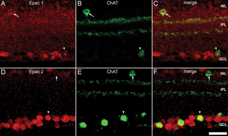
Figure 8.
Epac expression within the cholinergic amacrine cells. Double-labeling of Epac1 (A) and Epac2 (D) with ChAT (B, E). ChAT is expressed by cholinergic amacrine cells located at the INL and displaced cholinergic amacrine cells at the GCL and 2 strata within the IPL, an outer OFF cholinergic layer and an inner ON cholinergic layer. Epacs1 (C) colocalized with ChAT positive amacrine cells of both the INL (arrow) and GCL (arrowhead). Epac2 (F) was colocalized with ChAT positive cells of GCL (arrowhead), but demonstrated weak colocalization with ChAT cells of the INL (arrow). Scale bar = 25μm.

