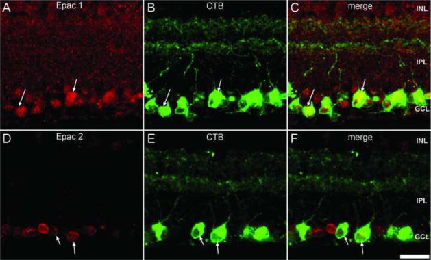
Figure 9.
Epac expression within the retinal ganglion cells. Double-labeling of Epac1 (A) and Epac2 (D) with CTB (B, E). Immunodetection of retrograde labeled retinal ganglion cells with CTB demonstrates expression within multiple retinal ganglion cell bodies and their dendrites within the IPL. Both Epacs (C, F) colocalized with retinal ganglion cell bodies (arrow). Scale bar = 25μm.

