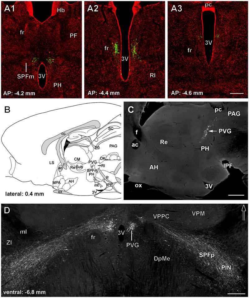
Fig. 2.
TIP39 neurons in the subparafascicular area of the rat posterior thalamus. A: TIP39-ir neurons (green) are shown in the PVG in coronal sections labeled with a fluorescent Nissl dye (red) at antero-posterior (AP) coordinates -4.2, -4.4, and -4.6 mm from the bregma level. B: The position of the PVG is shown in a drawing of a sagittal section of the rat brain at 0.4 mm lateral from the midline (Paxinos and Watson, 1998). C: A photomicrograph of a sagittal section corresponding to panel B shows the location of TIP39-ir neurons in the PVG. D: TIP39-ir neurons and fibers are shown in a horizontal section 6.8 mm ventral to the surface of the brain. The large density of TIP39-ir neurons in the PVG is in contrast to the scattered TIP39-ir neurons in the PIL while TIP39-ir fibers connect the 2 regions of TIP39 expression. Abbreviations: ac, anterior commissure; AH, anterior hypothalamic nucleus; cc, corpus callosum; CM, central median thalamic nucleus; Dk, nucleus of Darkschewitsch; DpMc, deep mesencephalic nucleus; f, fornix; fr, fasciculus retroflexus; IC, inferior colliculus; IPF, interpeduncular fossa; LS, lateral septal nucleus; ml, medial lemniscus; MPA, medial preoptic area; mt, mamillothalamic tract; ox, optic chiasm; PAG, periventricular gray; pc, posterior commissure; PF, parafascicular thalamic nucleus; PH, posterior hypothalamic area; PIN, posterior intralaminar thalamic nucleus; Pn, pontine nuclei; PVG, periventricular gray of the thalamus; py, pyramidal tract; Re, reuniens thalamic nucleus; RI, rostral interstitial nucleus of the medial longitudinal fasciculus; SC, superior colliculus; SPFm, magnocellular subparafascicular thalamic nucleus; SPFp, parvicellular subparafascicular thalamic nucleus; Sub, submedius thalamic nucleus; VPM, ventral posteromedial thalamic nucleus; VPPC, ventral posterior parvicellular thalamic nucleus; ZI, zona incerta; 3V, third ventricle; 4V, fourth ventricle. Scale bar = 500 μm for A and D, and 300 μm for C.

