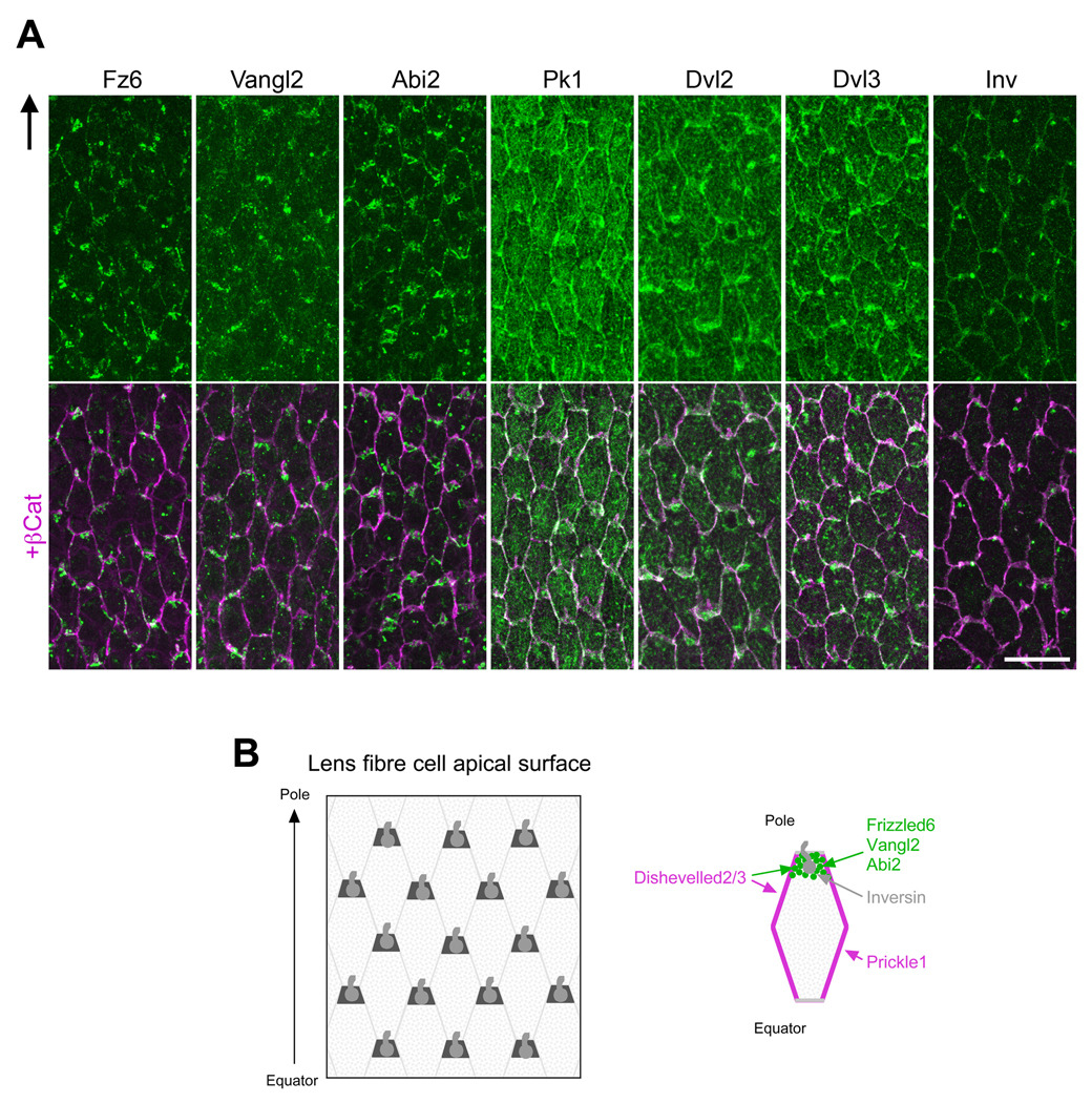
Fig 3. PCP proteins (green) are partitioned to cellular domains.
(A) At apical tips of OCF, Fz6, Vangl2 and Abi2 are predominantly associated with the short side of each cell proximal to anterior pole. In contrast, Pk1 is predominantly localised to the long sides. Dvl2/Dvl3 tend to be localised to all sides of each cell with some cells showing a little more intense reactivity associated with the short side proximal to anterior pole. β-catenin (purple) demarcates cell margins and co-localises with Pk1, Dvl2 and Dvl3. Arrow in (A) points to anterior pole. Scale bar, 20 µm. (B) Representation of the apical tips of fibers (left) and a single fiber (right). Abbreviations: Abi2, Abl-interactor-2; Dvl2/3, Dishevelled 2/3; Fz6, Frizzled 6; Inv, Inversin; OCF, outer cortical fibers; Pk1, Prickle 1; Vangl2, Van Gogh-like 2.

