
Figure 4. Role of ERRα in the insulin inhibition of the PDK4 gene.
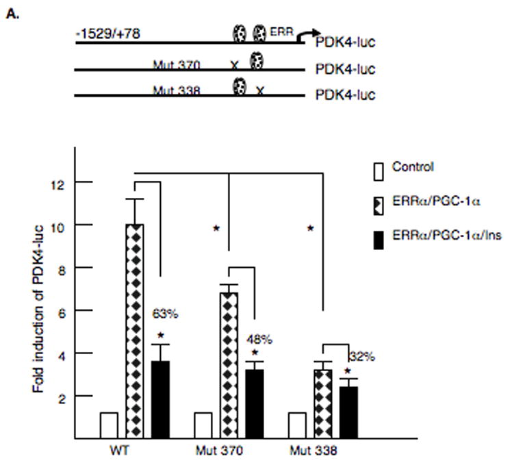

A. McA-RH7777 hepatoma cells were transfected with 2 μg of rat PDK4-luciferase reporter gene −1529/+78 region or rat PDK4-luciferase reporter gene −1529/+78 with the ERR sites mutated. A model of the mutated PDK4 promoters is shown. The expression vectors for ERRα (250 ng) and PGC-1α (1 μg) were cotransfected and TK-Renilla (0.2 μg) was included as a transfection control. Cells were harvested 36 h after transfection. The luciferase activity was corrected for protein content and Renilla activity. All transfections were done in duplicate and repeated three or four times. Insulin was used at a concentration of 100 nM and was added for 24 h. The data are expressed as the mean of the fold induction ± S.E. in the presence or absence of insulin. All transfections were performed in duplicate and repeated four times. The p< 0.01 is indicated by the * asterisk. B. A model of the PGC-1α mammalian expression vectors is shown. The −1529/+78 PDK4 luciferase reporter gene was transfected into McA-RH7777 hepatoma cells along with PGC-1α vectors where the L2 and L3 domains were altered. Luciferase assays were carried out after 40 h and values were normalized by protein content and Renilla activity. The luciferase activity observed following transfection with the mutant vectors was plotted as a percentage of the wild-type PGC-1α vector. The data are presented as the average percentage activation of luciferase ± S.E. All transfections were performed in duplicate and repeated four times.