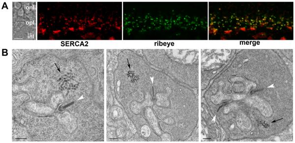
Figure 1. Rod terminals in the mouse retina contain an ER-like structure.
A. Confocal image showing an optical section of the mouse OPL double labeled for SERCA2 (red) and the synaptic ribbon protein, ribeye (green). In the merged image (right panel) areas of overlap appear yellow. B. Electron micrographs of mouse rod terminals. Synaptic ribbons are indicated with white arrowheads and putative ER with black arrows. The scale bars represent 5 μm in A and 200 nm in B. Abbreviations: onl, outer nuclear layer; opl, outer plexiform layer; inl, inner nuclear layer.

