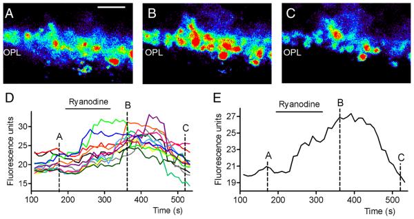
Fig. 4. Ca2+ increases in rod terminals evoked by stimulation of CICR with 10 μM ryanodine.
Pseudocolor images of a single confocal section of the outer plexiform layer loaded with Fluo4 in control conditions (A), after bath application of 10 μM ryanodine (B), and following washout (C). Bar: 10 μm. D. Fluo4 fluorescence changes measured from 12 terminals at 10 s interval. E. Average Fluo4 fluorescence changes from the same terminals. Images in A-C were obtained at the time points indicated in the graphs.

