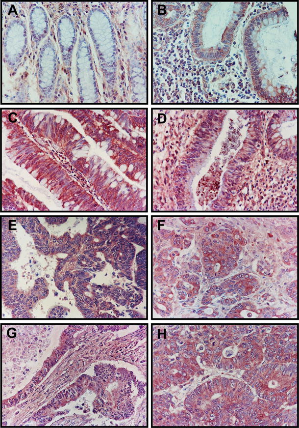
Figure 2. Immunohistochemical localization of RGC-32 in normal colonic tissue, colon precancerous lesions, and colon adenocarcinoma samples.
(A) Normal non-neoplastic mucosa from non-cancer cases, with mainly interstitial mesenchymal staining. (B) Small adenoma (maximal diameter, <2 cm). (C) Large adenoma (maximal dimension, >2 cm) showing more intensive staining than in the small adenoma. (D) Inflamed non-neoplastic mucosa from an ulcerative colitis patient, with moderate staining of the glands. (E) Primary invasive colon adenocarcinoma, stage T1/T2. (F) Primary invasive colon adenocarcinoma, stage T3/4. (G) Primary invasive colon adenocarcinoma metastatic to the lymph nodes, and (H) primary invasive colon adenocarcinoma metastatic to distant organs, with both (G) and (H) showing a very intense staining pattern.

