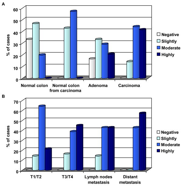
Figure 3. Staining score distribution for the expression of RGC-32.
A) Percentage of samples of normal mucosa, normal mucosa resected from colorectal adenocarcinoma, colonic adenomas, and adenocarcinomas that were positive for RGC-32 expression. Only 67% of normal colonic mucosa samples were positive, as compared to 100% of the normal mucosa samples from colons resected from colon adenocarcinomas. +; slightly positive, ++; positive, +++; highly positive.
B) RGC-32 staining percentages for early-stage colon cancer (T1 or T2), advanced-stage colon cancer (T3 or T4), colon adenocarcinoma metastasized to the lymph nodes, and colon adenocarcinoma metastatic to distant organs.

