Fig. 1.
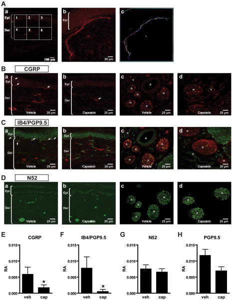
Effects of dilute capsaicin infiltration on afferent fiber immunoreactivity. A: Example for measuring immunoreactive area in skin sections. (Aa): Confocal image of a skin section for calcitonin gene related peptide (CGRP) fluorescent immunohistochemistry. Six adjacent squares (300 μm × 300 μm for each) were selected bordering the epidermis and dermis. (Ab,c): In each square, the total immunopositive area was determined by manually tracing CGRP-immunoreactive fibers. The relative area (RA) of immunoreactivity was calculated by dividing the total immunopositive area by the total skin area. B: CGRP fluorescent immunohistochemistry in plantar skin (Ba,b) and subcutaneous tissue containing large nerve bundles (Bc,d) 1 week after subcutaneous infiltration of vehicle versus 0.05% capsaicin. CGRP-immunolabeled fibers in plantar skin are indicated by arrows (Ba,b). C: Isolectin B4 (IB4) and protein gene product 9.5 (PGP9.5) fluorescent double labeling immunohistochemistry in plantar skin (Ca,b) and subcutaneous tissue (Cc,d) after vehicle versus capsaicin treatment. Double-labeled fibers (yellow) in the epidermal-dermal junction are indicated by arrows (Ca). D: Neurofilament 200 (N52) fluorescent immunohistochemistry in plantar skin (Da,b) and subcutaneous tissue (Dc,d) after vehicle vs. capsaicin treatment. E-H: The RA of immunoreactivity of CGRP- (E), IB4/PGP9.5- (F), N52- (G), and total PGP9.5-positive fibers (H) in vehicle-treated and 0.05% capsaicin-treated skin. Data are expressed as mean ± SE. *P < 0.05 vs. vehicle by unpaired t-test. Epi = epidermi; Der = dermis; n = nerve; a = artery; v = vein; veh = vehicle; cap = capsaicin.

