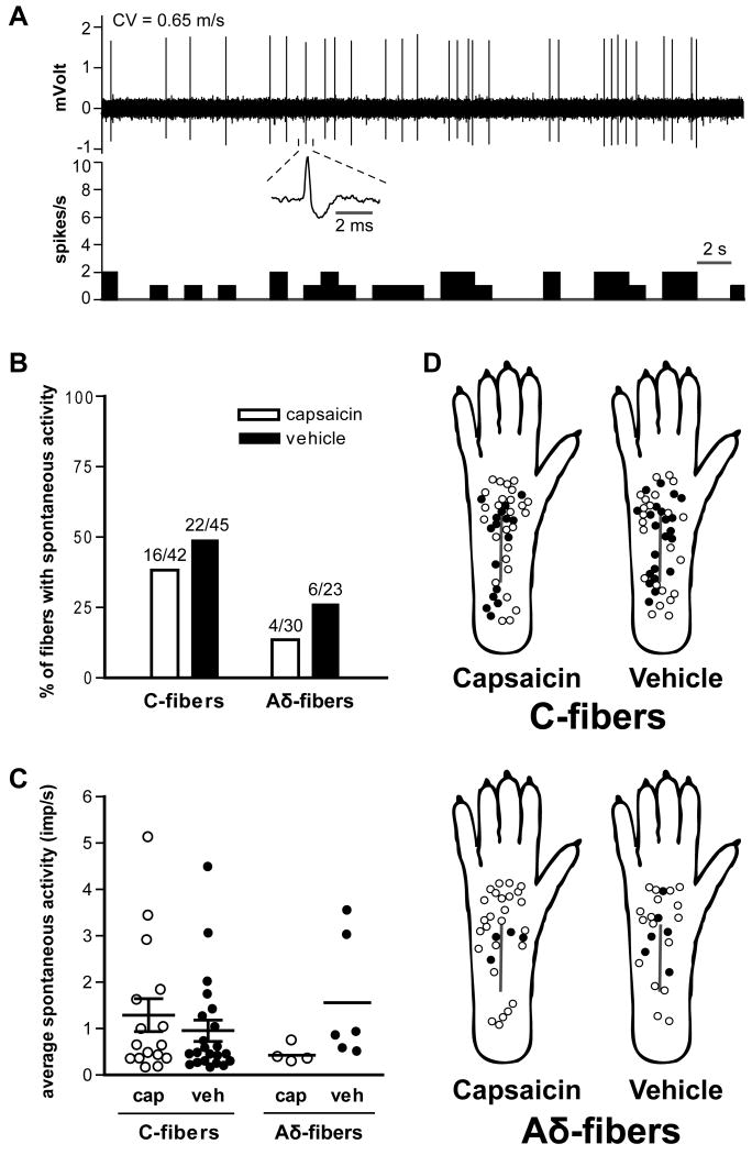
Fig. 4.
Effect of capsaicin treatment on spontaneous activity of mechanosensitive afferent fibers one day after plantar incision. A: A sample recording of ongoing activity in a C-fiber from a capsaicin-treated rat. The average firing frequency in this example was 0.76 imp/s. The upper and lower panels show the digitized oscilloscope trace and spike density histogram (bin width = 1 s), respectively. Inset displays an enlarged view of the action potential. CV = conduction velocity. B: Prevalence of spontaneously discharge in C- and Aδ-fibers. C: Average activity of spontaneously discharging fibers. Bars represent mean and whiskers represent SE. Imp = impulse; cap = capsaicin; veh = vehicle. D: Distribution of the receptive fields of C- and Aδ-fibers with or without spontaneous activity, for capsaicin- and vehicle-treated rats. Solid circles represent receptive fields of afferent units with spontaneous activity and open circles represent those without spontaneous activity.

