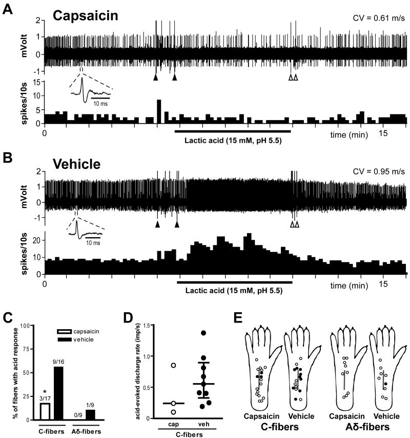
Fig. 7.
Effect of capsaicin treatment on the acid-responsiveness of afferent units one day after plantar incision. A and B: Sample recordings from two single C-fibers from a capsaicin- (A) and a vehicle-treated rat (B). The upper and lower panels show the digitized oscilloscope tracings and spike density histograms (bin width = 10 s), respectively. Insets display the action potentials of these units. Artifacts produced by placing and removal of the metal ring are marked by two black arrowheads and two white arrowheads, respectively. CV = conduction velocity. C: Percentage occurrence of acid-responsive units in C- and Aδ-fibers when tested with pH 5.5 lactic acid. * P < 0.05 vs. vehicle by χ2 test. D: acid-evoked discharge rate of each acid-responsive C-fiber during lactic acid application. Bars represent median and whiskers represent interquartile range. Imp = impulse; cap = capsaicin; veh = vehicle. E: Distribution of the receptive fields of C- and Aδ-fibers with or without responsiveness to pH 5.5 lactic acid, for capsaicin- and vehicle-treated rats. Each circle represents the center of a unit's mechanoreceptive field. Solid and open circles represent receptive fields of afferents with and without acid-responsiveness, respectively.

