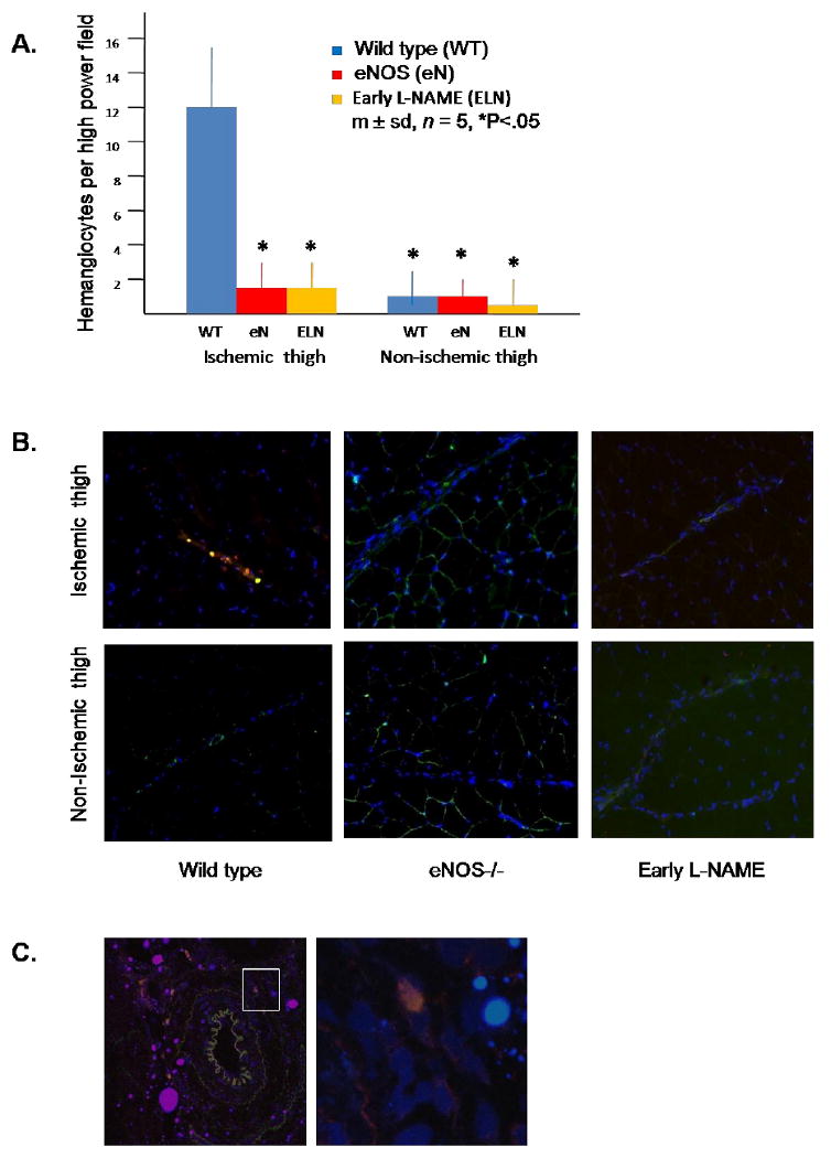
Figure 3. Presence of hemangiocytes in the thigh musculature in wild type, eNOS-/-, and early L-NAME treatment groups on day 3 after induction of hindlimb ischemia.

(A) Quantitation of hemangiocytes. Hemangiocytes were defined as cells that co-localized CXCR4 and VEGFR1. Data for each mouse was determined as the average number of hemangiocytes present in 10 randomly selected high power fields, determined by a blinded observer. (B) Representative photomicrographs of hemangiocyte immunostaining. VEGFR1 is stained green, CXCR4 is stained red, and nuclei are stained blue. A collateral vessel is present in each photomicrograph, evidenced by the linear cord of blue stained nuclei. Note the present of double stained hemangiocytes clustered along the collateral artery in the ischemic thigh of the control mouse (200×). (C) Confocal microscopy of a collateral artery within the ischemic thigh of the wild type group. The left hand photo is shown at 200× magnification. The area delineated by the white square in the left hand photo is shown at 400× in the right hand photo. Note the presence of a hemangiocyte in the adventitial area of a collateral artery. This adventitial distribution of hemangiocytes was commonly observed in the ischemic thigh muscle of wild type mice, but was never observed in eNOS-/- or early L-NAME treatment groups.
